Monday 24 September
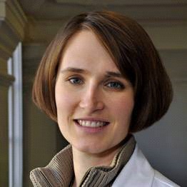
Interview with Dr Lynette Sholl, Associate Professor, Pathology, Harvard Medical School, Associate Pathologist, Pathology, Brigham And Women’s Hospital

Dr Sholl is a graduate of Johns Hopkins University and Stanford University School of Medicine.
Her research focuses on identifying markers that will improve the classification of lung cancer, providing predictive information regarding therapy, and more precise prognostic information. She has a special interest in understanding the genomic modifiers of tumour behaviour, morphology, and response to treatment.
What first motivated you to work in lung cancer treatment?
It was really the timing of my training that led me into lung cancer studies. I finished medical school in 2003, right about the time that TKIs (targeted kinase inhibitors) were coming to the fore and there was a lot of excitement around EGFR and the potential to target different tumour types.
Personally, seeing one lung cancer patient in particular in the ICU – a woman of Asian descent who had never smoked – responding to TKIs, brought to life the way those new types of medicine could improve outcomes for people with lung cancer.
I then had the privilege to train with some of the leading lights in pathology during my second-year residency in Boston. Pathology struck me as a unique way to think about disease mechanisms and visualise in situ disease at the cellular level. I find it thrilling to look down the scope and see complex biological processes at work in high detail.
What is different about your role today from when you started your career?
The principle difference is that we now know more about the molecular underpinnings of cancer. In terms of our molecular understanding, we were at the tip of the iceberg in 2004.
Now, we have cast new light on the variations between cancers and made more sense of the diversity of tumours. Improved molecular understanding has improved the accuracy of tumour classification, added nuance to diagnostics and increased the specificity of diagnoses.
What have been the biggest advances in pathology?
Essentially, it’s about sequencing. We are now able to do massive amounts of sequencing with very small amounts of tissue. This allows us to engage in discovery during routine diagnostics and to support the use of targeted therapies on the basis of mutations like EGFR, B-RAF and ALK.
This information enhances traditional staging and grade information to better inform treatment decisions and to make more judgments about how we expect patients to respond to treatment.
What do you see as the most significant challenges in your field?
The main challenge is educating both the profession and the population about genomics. For instance, the IASLC/CAP/AMP biomarker guidelines identify only a few genes that require routine testing. We are still really only scratching the surface in terms of using genomics in treatment decisions. Efficacy of treatment is still only registered in a subset of patients.
The medical school education scheme will need to shift to incorporate genomic analysis. For example, today there’s still limited understanding amongst clinicians about the difference between germline and somatic mutations and the implication this has in terms of treatment decisions.
As genomic medicine is used more and more in routine care we need to be thinking now about how we can fit genomics into the curriculum.
What are you most excited about in your field?
One of the most exciting prospects is the opportunity to combine a number of new technologies and testing techniques. By synthesizing a large number of biomarkers and using emerging technologies like liquid biopsies, we have the potential to develop even more sophisticated approaches to staging and diagnosis.
Liquid and genetic profiling could bring together all the most powerful diagnostic technologies to deliver the most accurate diagnoses yet.
More powerful visual technology, married to advances in computational technology, also have the potential to transform pathology by allowing us to use a host of new algorithmic tools and analytics to better understand disease.
In sum, we’re seeing the emergence of powerful prognostic models embracing genetics and new biomarkers that will provide better diagnostic tools so that we can further stratify patient populations.
What role do you see international collaboration playing in advances in lung cancer treatment?
International collaboration helps the field to advance faster, as opposed to working in an insular fashion. I believe it is critical that we speak about launching biomarker testing and what this means at an international level. It is crucial that we think carefully about how we prepare health systems across the globe to deliver these new approaches to diagnosis, staging and stratification.
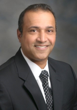
Professor George Eapen, Deputy Department Chair and Professor of the Department of Pulmonary Medicine, Division of Internal Medicine at the University of Texas MD Anderson Cancer Centre.

Professor Eapen completed his training at the University of Benin before undertaking his residency at the Southern Illinois School of Medicine and a clinical fellowship at Baylor College of Medicine, Houston, Texas.
In addition to his position at the Department of Pulmonary Medicine, Professor Eapen is Section Chief of Interventional Pulmonology, University of Texas. He is also a member of American College of Chest Physicians and the President-Elect of the American Association for Bronchology and Interventional Pulmonology.
What first motivated you to work in pulmonary medicine and lung cancer?
When I started working 20 years ago, attitudes to lung cancer were negative and nihilistic. A diagnosis was tantamount to a death sentence and there was an undercurrent of victim blaming – a sense that lung cancer was self-inflicted because it is often caused by smoking. I always felt this was unfair and that this neglected patient population deserved more attention and care. This attracted me to practice in lung cancer.
I was then drawn to interventional pulmonology because it was an optimistic speciality, looking for solutions. Interventional pulmonologists were exploring new possibilities in treating lung cancer, experimenting with new technologies and techniques like stents or rigid bronchoscopy. It was an energetic field and I found this very appealing.
What is different about your role today from when you started your career?
The role of the interventional pulmonologist has morphed and expanded as new technologies have come through. When surgery was the only curative treatment available, we used to be confined to the pre-operation assessment, working out whether a patient could cope with surgery.
Today, we have a major footprint in diagnosis and staging, with the use of less-invasive procedures like endobronchial ultrasound (EBUS). Our options for curative treatment have expanded beyond surgery to less invasive procedures. We also have better techniques and tools for symptom management, so that gives us a bigger role in patient care.
Secondly, we have seen a decisive shift toward multidisciplinary care. 20 years ago, patients were treated in silos. Now, with MDTs we have a one-stop shop to discuss individual patients’ needs and bring together different perspectives and expertise to enhance their care. This is a very positive development.
What have been the biggest advances in your specialty, in terms of how we have improved outcomes for people with lung cancer?
Change is often incremental, and small steps add up to a big impact. One of the biggest advances is that we now know that lung cancer screening can provide a mortality benefit. If we can shift from two thirds of people being diagnosed with a late-stage cancer to two thirds being diagnosed with an early-stage cancer, we can increase survival rates.
In staging, EBUS has been a major step forward: a minimally invasive way of getting the right information, with a better safety profile than surgery, increasing the numbers of patients who have staging information.
In terms of treatment, stereotactic ablative radiotherapy (SABR), is becoming more mainstream. We’re seeing more patients with controlled disease at three years – almost equivalent to lobectomy.
What do you see as the most significant challenges to continuing to improve outcomes for people with lung cancer in the coming years?
The key issues are cost and accessibility. The cost of developing and then rolling-out new treatments is high. Equally, geographic access to high-quality and appropriate care is another challenge. It is no use building a better mouse trap if you don’t put it where the mice are.
Some patients live far away from expert centres. Since we can’t put a centre in every town, we need to find ways for professionals to disseminate their expertise to expand access. We must aim to deliver the greatest amount of care to the greatest number of people.
What are you most excited about in your field?
Immunotherapies have changed how we approach lung cancer treatment. Consider the past: we dealt only in crude terms of small cell or non-small cell lung cancer. Now we have identified over 23 actionable mutations and treating the right target with the right drug has resulted in big improvements in progression-free survival.
SABR is also very promising. If we can move towards more non-surgical approaches to lung cancer treatment, that would be very exciting. From my perspective as an interventional pulmonologist I find transbronchial biopsy and ablation the most exciting development. It’s early days but the fact that resources are being moved in this direction fills me with optimism.
What role do you see international collaboration playing in advances in lung cancer treatment?
I believe science is a team sport. As Isaac Newton said, if we see further, it is because we stand on the shoulders of giants. Collaboration means we can advance science and get better outcomes for patients. We can always improve on what we knew or had before.


